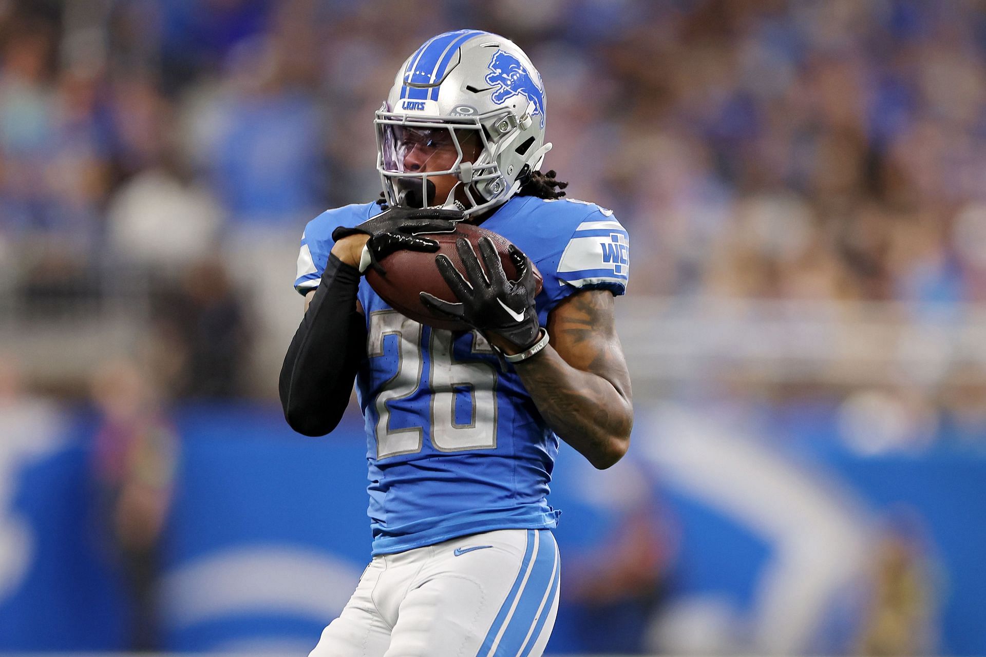
Understanding Gibbs Injuries

A Gibbs injury, also known as a Gibbs fracture, is a specific type of fracture affecting the lateral malleolus of the ankle. It is characterized by a fracture that extends from the tip of the lateral malleolus, the bony prominence on the outer side of the ankle, towards the joint surface. This fracture pattern is distinct from other ankle fractures and requires specific treatment strategies.
Mechanism of Injury
Gibbs fractures typically occur due to a forceful inversion and dorsiflexion of the ankle joint. This mechanism of injury is common in activities involving sudden twisting or landing on an uneven surface. During inversion, the foot turns inwards, while dorsiflexion refers to the upward bending of the foot towards the shin. This combined motion puts significant stress on the lateral malleolus, leading to a fracture.
Anatomical Structures Involved
The lateral malleolus, a prominent bony projection on the fibula (the lower leg bone), plays a crucial role in stabilizing the ankle joint. The lateral malleolus forms the outer wall of the ankle joint, providing support and limiting excessive inward movement of the foot. When a Gibbs fracture occurs, it disrupts the integrity of the lateral malleolus, compromising the stability of the ankle joint.
Symptoms of a Gibbs Injury
Individuals with a Gibbs fracture typically experience a range of symptoms, including:
- Severe pain and tenderness around the lateral malleolus
- Swelling and bruising in the ankle area
- Difficulty bearing weight on the affected ankle
- Instability or a feeling of giving way in the ankle
- Deformity or visible displacement of the lateral malleolus
Diagnosis and Treatment: Gibbs Injury

Diagnosing and treating a Gibbs injury requires a comprehensive approach, considering the unique nature of this complex condition. The diagnostic process involves a combination of clinical evaluation, imaging studies, and, in some cases, further specialized investigations. Treatment options range from conservative measures to surgical interventions, tailored to the severity of the injury and individual patient factors.
Diagnostic Procedures, Gibbs injury
The diagnosis of a Gibbs injury typically begins with a detailed medical history and physical examination. The clinician will inquire about the mechanism of injury, the patient’s symptoms, and any relevant past medical history. Physical examination will focus on assessing the range of motion, stability, and tenderness of the affected joint.
- Imaging Studies: Imaging plays a crucial role in confirming the diagnosis and determining the extent of the injury.
- X-rays: These are often the initial imaging modality used, providing basic information about the bone alignment and any fractures. However, X-rays may not always clearly visualize ligamentous injuries, which are often associated with Gibbs injuries.
- Magnetic Resonance Imaging (MRI): MRI is considered the gold standard for evaluating ligamentous injuries, providing detailed images of soft tissues, including ligaments, tendons, and cartilage. It can help identify tears, sprains, and other soft tissue damage that may be associated with a Gibbs injury.
- Computed Tomography (CT) Scan: CT scans can provide detailed images of bones and can be used to assess for fractures, particularly complex fractures that may involve multiple bones.
- Arthroscopy: In some cases, an arthroscopic examination may be necessary to provide a more definitive diagnosis. Arthroscopy involves inserting a small camera and surgical instruments into the joint, allowing for direct visualization of the injured structures. This procedure can also be used to perform certain surgical repairs if needed.
Treatment Options
Treatment for a Gibbs injury aims to restore joint stability, reduce pain, and improve function. The approach to treatment will vary depending on the severity of the injury and the individual patient’s needs and goals.
- Conservative Treatment: Conservative treatment options are often the first line of treatment for Gibbs injuries, particularly for less severe cases. These approaches aim to reduce pain and inflammation, promote healing, and restore joint function.
- Rest, Ice, Compression, and Elevation (RICE): This is a standard treatment for acute injuries, helping to reduce pain and swelling.
- Immobilization: Depending on the severity of the injury, a splint, brace, or sling may be used to immobilize the joint and prevent further damage.
- Pain Medication: Over-the-counter pain relievers, such as ibuprofen or acetaminophen, can help manage pain and inflammation. In some cases, stronger pain medications may be prescribed.
- Physical Therapy: Physical therapy plays a crucial role in rehabilitation after a Gibbs injury. It aims to improve range of motion, strength, and stability of the affected joint, as well as to teach proper body mechanics and exercise techniques to prevent future injuries.
- Surgical Intervention: Surgical intervention may be considered for more severe Gibbs injuries, particularly those involving significant ligamentous damage or instability.
- Ligament Repair or Reconstruction: Surgical repair involves suturing the torn ligament back together, while reconstruction involves using a graft to replace the damaged ligament. The type of surgery will depend on the specific ligament involved and the extent of the injury.
- Arthroscopic Surgery: Many ligament repair and reconstruction procedures can be performed arthroscopically, which involves making small incisions and using a camera and surgical instruments to repair the injury.
- Open Surgery: In some cases, open surgery may be necessary to access the injured ligament or to perform more complex repairs.
Comparison of Conservative and Surgical Treatment
Conservative treatment is often the preferred approach for less severe Gibbs injuries, as it offers a less invasive and potentially faster recovery time. However, it may not be effective for all injuries, and if conservative treatment fails to provide adequate pain relief or stability, surgical intervention may be necessary.
Surgical intervention is typically reserved for more severe injuries, those involving significant ligamentous damage, or those that have not responded to conservative treatment. While surgery can provide a more definitive solution for restoring joint stability, it comes with risks and a longer recovery period.
| Factor | Conservative Treatment | Surgical Intervention |
|---|---|---|
| Invasiveness | Less invasive | More invasive |
| Recovery Time | Generally shorter | Generally longer |
| Success Rate | Variable, depends on the severity of the injury | Generally higher for severe injuries |
| Risks | Lower risk of complications | Higher risk of complications, such as infection, bleeding, and nerve damage |
| Cost | Generally less expensive | Generally more expensive |
Potential Complications
Gibbs injuries and their treatments can be associated with potential complications, which may vary depending on the severity of the injury, the treatment approach, and individual patient factors.
- Infection: Infection is a potential complication of any surgical procedure, but it can also occur with conservative treatment if the injury becomes contaminated.
- Bleeding: Excessive bleeding can occur during surgery, particularly if there are pre-existing clotting disorders.
- Nerve Damage: Nerve damage can occur during surgery, particularly in the area of the shoulder or elbow, which can lead to numbness, tingling, or weakness.
- Joint Stiffness: Joint stiffness can occur after a Gibbs injury, especially if the joint is immobilized for an extended period.
- Osteoarthritis: Osteoarthritis is a long-term condition that can develop after a Gibbs injury, particularly if the injury is severe or if there is significant damage to the joint cartilage.
Rehabilitation and Recovery

Recovering from a Gibbs fracture requires a comprehensive rehabilitation program that focuses on restoring function, reducing pain, and preventing further complications. This process involves a multidisciplinary approach, with physical therapists, occupational therapists, and sometimes pain management specialists working together to guide the patient’s recovery.
Physiotherapy and Occupational Therapy
Physiotherapy plays a crucial role in regaining mobility and strength after a Gibbs fracture. It involves a series of exercises designed to improve range of motion, reduce swelling, and strengthen the muscles surrounding the injured area. This may include:
- Passive range of motion exercises: These exercises are performed by the therapist to move the joint through its full range of motion, preventing stiffness and promoting blood flow.
- Active range of motion exercises: Once the pain subsides, patients are encouraged to perform active range of motion exercises, where they move the joint themselves.
- Strengthening exercises: These exercises aim to rebuild muscle strength and endurance, which are essential for returning to daily activities.
- Proprioceptive exercises: These exercises help improve balance, coordination, and body awareness, enhancing stability and reducing the risk of re-injury.
Occupational therapy focuses on improving daily living skills and returning to work or other activities. This may involve:
- Adaptive equipment training: Occupational therapists may teach patients how to use adaptive equipment, such as assistive devices for dressing, bathing, or cooking, to help them perform daily tasks with ease.
- Work hardening programs: These programs help individuals gradually return to work by simulating work-related activities and building up stamina.
- Ergonomic assessments: Occupational therapists can evaluate the work environment and suggest modifications to reduce strain on the injured area.
Recovery Timeline
The recovery timeline for a Gibbs fracture varies depending on the severity of the injury and the individual’s overall health. Generally, the healing process can take several weeks to months.
- Non-operative treatment: Patients with a non-operative Gibbs fracture may experience pain and swelling for several weeks. They may be able to return to normal activities within a few months, with full recovery taking up to six months.
- Operative treatment: Patients who underwent surgery for a Gibbs fracture may experience a longer recovery period. The healing process can take several months, with full recovery taking up to a year.
Preventing Future Gibbs Injuries
While a Gibbs fracture can occur due to various factors, certain measures can be taken to reduce the risk of future injuries:
- Strengthening the surrounding muscles: Regular exercise, particularly focusing on the muscles of the shoulder, neck, and upper back, can improve stability and reduce the likelihood of injury.
- Proper lifting techniques: Using correct lifting techniques, such as bending the knees and keeping the back straight, can minimize stress on the shoulder joint.
- Maintaining a healthy weight: Obesity can put additional strain on the shoulder joint, increasing the risk of injury.
- Avoiding high-impact activities: Individuals with a history of Gibbs fractures should avoid activities that place excessive stress on the shoulder joint, such as contact sports or heavy lifting.
Gibbs injury – So, Gibbs is down, bummer, right? But hey, don’t sweat it too much, the Vikings are known for their depth, and it’s always good to have a backup plan. You can check out the vikings depth chart to see who might be stepping up.
Who knows, maybe this injury opens the door for a new star to shine!
Man, that Gibbs injury is a bummer, right? Remember when Justin Jefferson went down with that ankle injury in college? It felt like the whole world stopped for a minute, but he came back stronger than ever. Hopefully, Gibbs can do the same!
